VIRTUAL DISSECTION / SKELETAL SYSTEM
Inside a bone.
Follow the femur from the body to the tray. Then discover what is inside.
Self-guided lab · Put the pieces togetherStart with the 3–2–1 exploration below. Record observations in a notebook or use the optional on-page notes. This is practice, not a test.
Before you begin
Predict: Is a long bone solid all the way through? Write your prediction before exploring, then return to it after inspecting the bone.
If sharing a device: Take turns controlling the model. You may also work independently. Each person records their own evidence and explanations.
Learning goal: Compare compact bone, spongy bone, red marrow, and yellow marrow; connect bone names, divisions, shapes, and functions.
Language goal: Explain “I observed ____. This structure helps ____ because ____.”
Independent study or classroom use
Learning on your own: no class enrollment or teacher is needed. Follow the controls, compare the four internal structures, and use the references and lab questions to check your understanding. A notebook or the optional on-page notes can hold your observations.
Classroom journal pathway: the 45-minute guide and narrated classroom walkthrough below are retained for learners using a composition journal. If assigned by a teacher, follow their report, review, and submission directions. Any class test or deadline is separate from this self-paced public lab.
SELF-PACED · 45-MINUTE GUIDE
Your journal is your lab report.
Read one step, do it, then choose Next step. Work independently; if you share a device, alternate controls. Everyone writes their own report. No copies or duplicate online answers.
0–5 min · SECTION 1 · Exploration
Explore first
- Open the specimen below. Try the available controls and follow the tool order on screen. You do not need scientific names yet.
- In your own composition journal, write 3 things you notice, 2 interesting details, and 1 question. Do this before the walkthrough or sample answers.
Before moving on: I wrote my own 3–2–1 observations.
Times are suggestions, not a countdown. You can go back anytime. Do the exploration and prediction before viewing journal examples.
Watch the 2-minute student walkthrough
See the body-to-tray sequence, the four tool movements, and how to record your evidence. Pause or replay whenever you need.
Classroom simulation · Edited screenshots of this lab · Synthetic narration. The prepared specimen simplifies anatomy and skips surgical preparation. Work only on screen.
Read the full transcript
- 0:00
Welcome to the skeletal lab, an edited classroom simulation. Work only on screen.
- 0:07
If earlier progress appears, choose Reset specimen. Your notebook answers are kept.
- 0:13
First, predict what is inside a long bone. Then choose Move muscles aside.
- 0:20
Choose Lift femur onto tray, or drag across the specimen. Watch the same femur leave its tissue bed and move onto the tray.
- 0:30
Choose the Begin dissection button.
- 0:34
The scalpel is selected for you. Press and drag anywhere across the bone to open the thin covering. No precision tracing is needed.
- 0:44
Next, drag with the forceps. The thin outer membrane is removed, revealing compact bone.
- 0:52
Now drag with the powered bone saw. Its small oscillating blade follows the outlined window in the hard compact bone.
- 1:02
Use the forceps again. Drag to lift the cut section as one solid piece. Hard bone does not peel.
- 1:11
Select all four structures. Read each observation and function card below the model.
- 1:18
Compact bone forms the dense, strong outer wall.
- 1:23
Spongy bone is hard and branching, not a soft sponge.
- 1:28
Red marrow produces blood cells within some of the spaces.
- 1:33
Yellow marrow in the shaft cavity stores fat.
- 1:38
Use the numbered notebook prompts. Explain your observations with your class notes. Apply what you learned to the helmet, backpack, and kicking a ball questions.
- 1:50
Download or print your notes. Answers stay on this device, with no automatic submission. Follow your teacher’s directions to turn them in.
Find the femur in the thigh
The femur is a long bone in the appendicular skeleton. Muscles move it; tendons connect muscle to bone; ligaments connect bone to bone at joints.
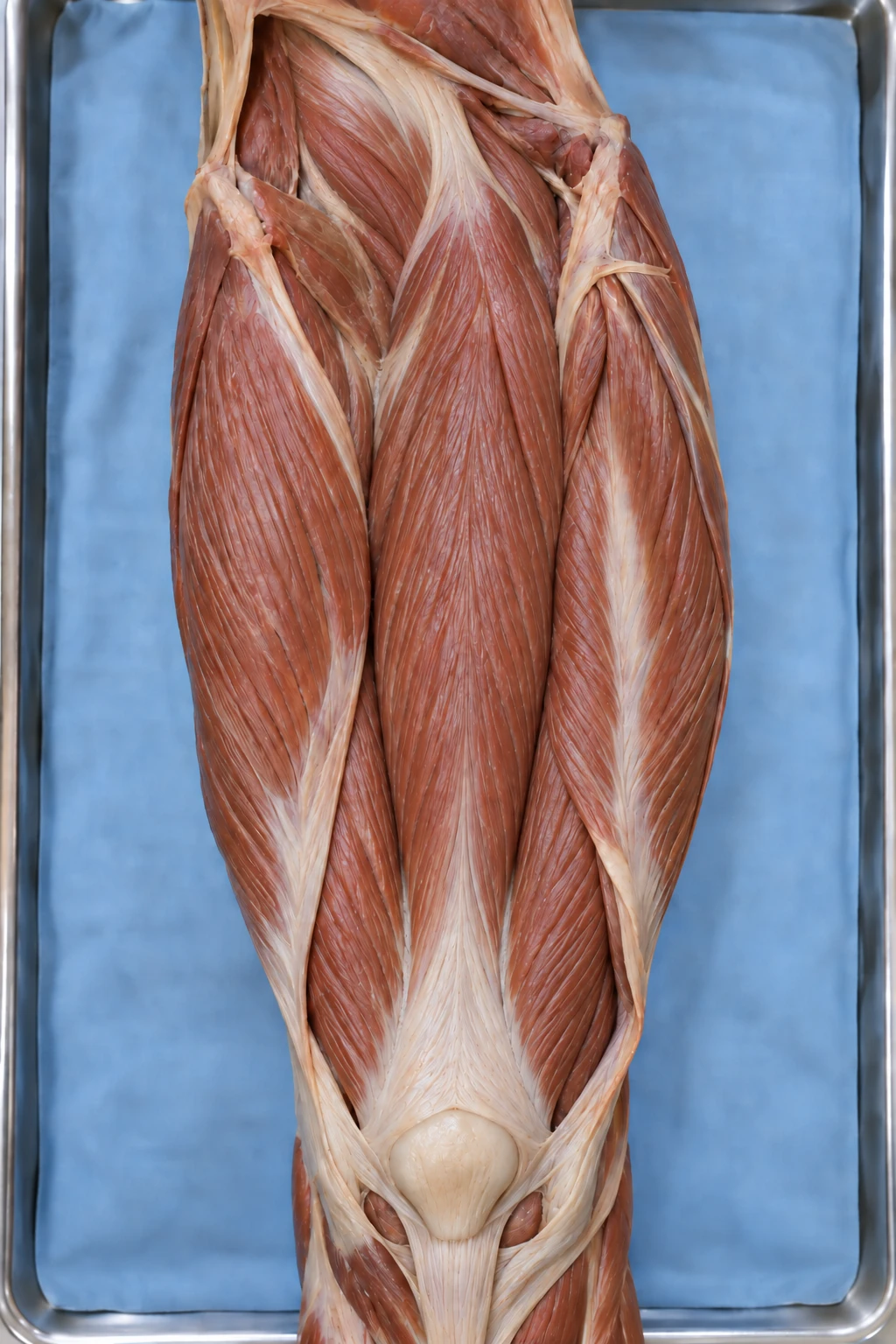
Classroom simplification: surrounding attachments are already released. The animation shows a prepared femur lifting out of its tissue bed and moving onto the tray, not a complete surgical procedure.
CONNECT THE DETAILS
From one bone to a whole system.
Use these references while completing the helmet, backpack, and kicking-a-ball scenarios.
Two skeletal divisions
Axial: skull, vertebral column, ribs, and sternum.
Appendicular: limbs and the shoulder and pelvic girdles that connect them to the body.
One everyday action can involve both divisions. Name the specific bone you are discussing.
Four bone shapes in this review
Long: longer than wide, such as a femur or tibia.
Short: roughly cube-shaped, such as a wrist carpal.
Flat: thin and often curved, such as a cranial bone.
Irregular: a complex shape, such as a vertebra.
These describe whole-bone shape. Compact and spongy describe bone tissue.
Functions to connect
Support · Protection · Movement · Blood-cell production · Storage of minerals and fat
Bones store minerals such as calcium and phosphate. Yellow marrow stores fat. Red marrow produces blood cells. Muscles pull on bones to move them at joints.
Check your understanding
- Identify the four internal structures without relying on color alone
- Explain why spongy bone is not soft sponge
- Distinguish blood-cell production from fat storage
- Name a bone, division, shape, and function for each everyday scenario
- Check your explanations against the science references; use class discussion too if available
About this model & science sources
The specimen images are AI-generated anatomical approximations, not photographs of human tissue. They show a prepared teaching specimen with an enlarged window. The optional 3D view is a simplified geometric model, not a human scan or a surgical trainer. The body-context opening shows a prepared femur lifting out of the surrounding tissue and moving continuously onto a tray. Attachments are already released; this is not a complete bone-removal procedure. The outer covering (periosteum), guided tool movements, and localized cortical window are enlarged and simplified to make the four focus structures visible. Compact bone is rigid; it is cut with a virtual powered oscillating bone saw and moved as a rigid section. Only the outer membrane is peeled. The small powered saw tip moves back and forth; it does not spin. The bone geometry and enlarged opening are teaching simplifications.
The red region illustrates active red marrow within some spongy-bone spaces near an end. Marrow distribution varies with bone and age. In adults, active red marrow is concentrated in selected bones, including the skull, vertebrae, sternum, pelvis, and upper ends of the femur and humerus. The yellow shaft marrow illustrates fat storage. Color is a teaching aid. The real tissues contain living cells and blood vessels.
The model is not to scale and does not show every bone structure. Follow the on-screen actions only; do not try them on people, animals, or real specimens.
Skeletal dissection · Individual evidence record
Progress: Open the thin outer covering
Before you open the bone
Predict: Is the inside of a long bone solid all the way through? What might you find inside?
________________________________________________________________________________
Observe the two bone tissues
How do compact and spongy bone differ in appearance? Explain how one visible feature of each helps it do its job.
________________________________________________________________________________
Distinguish the two kinds of marrow
Where did you find red and yellow marrow in this model? What is the main function of each?
________________________________________________________________________________
Apply it: bicycle helmet
Name a bone protected by a bicycle helmet. Give its skeletal division, its shape category, and the skeletal function involved.
________________________________________________________________________________
Apply it: carrying a backpack
Name a bone involved in supporting a backpack. Identify its skeletal division and shape, then explain the function it performs.
________________________________________________________________________________
Apply it: kicking a ball
Choose a bone involved in kicking a ball. Identify its skeletal division and shape. Explain how bones and muscles work together for movement.
________________________________________________________________________________
Put the pieces together
Revise your opening prediction. Explain how the outer wall, internal network, and marrow make a bone more than a solid support.
________________________________________________________________________________